Bridge Model of the Bearing Apparatus
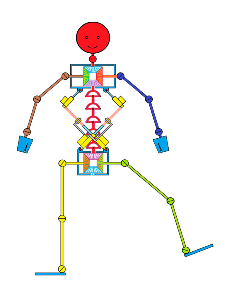
The important process that happens during the locomotion of the human body, can be observed in the axial organ. It is the interplay between the pelvic girdle and ribcage with the scapulae.
The biomechanics of the locomotive motor skills of the human musculoskeletal apparatus show obvious resemblances to bridge architectonics. To convey the body on four or two limbs, it is necessary to create the conditions for the shift of the centre of gravity of the body first. It can be shifted in the gravitational field only so that it was borne beyond the supporting base or supporting points. In mammals, the limbs are used to this load-bearing role. In most cases all four limbs play a part. Rarely, as in the case of apes, it is only two.
-
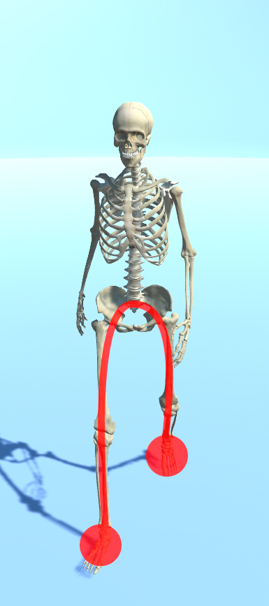
Illustration of the supporting point and the bearing arch in gait
-
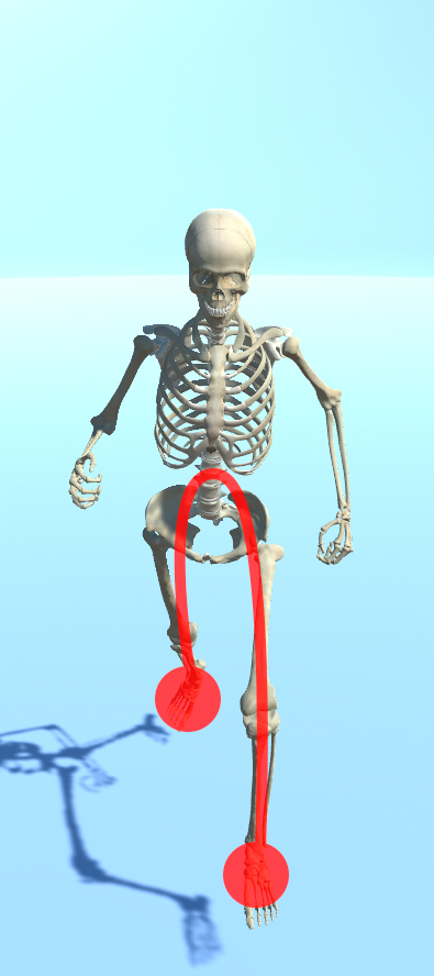
Illustration of the supporting point and the bearing arch in gait
The biomechanical construction of the human body that enables the upright gait on two limbs is the model of the most taxing type of locomotion of all so far known. Walking on two limbs is advantageous for several reasons. It’s most advantageous in terms of economical demands because as it “pulls” the centre of gravity of the body forward, it uses the kinetic energy of oscillation of the stepping lower limb as well as the oscillation of the “virtually” stepping upper limb. It can change the velocity and the direction of the motion very easily. It permits movement on various types of surfaces and accommodates changes in the terrain slope. An upright gait improves the conditions for visual and acoustic orientation.
-
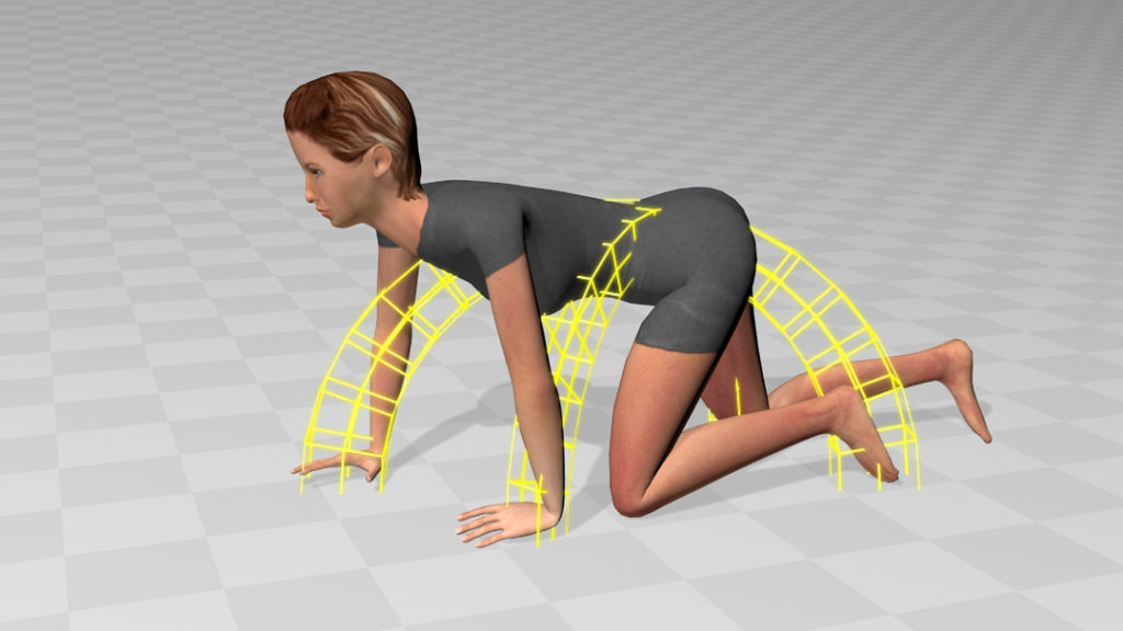
Illustration of changes in construction of the “bridge arch” and its changes during locomotion
-
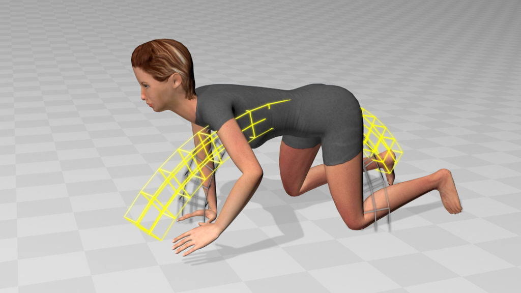
Illustration of changes in construction of the “bridge arch” and its changes during locomotion
-
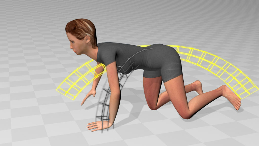
Illustration of changes in construction of the “bridge arch” and its changes during locomotion
On the other hand, upright gait bears many disadvantages and complications. It entails difficult conditions for hydrodynamics of the blood and lymphatic circulation; supportive valves in vessels had to develop to help the venous blood and lymph to return from the lower limb. Acral parts of the limbs suffer from swelling during their insufficient activity. Load on the bearing joints of the lower limbs leads to extreme stress, which is transmitted on the rather small surfaces of the joint cartilages of the hip, knee and ankle joints. They tend to be the source of many pathological changes with subsequent impacts on the gait biomechanics and posture of the body. The length of the long bones necessary for a sufficiently long step (so that the gait would be economical at all) results in susceptibility to fractures. Vertical position of the pelvis is also stressful for the function of the lumbar vertebrae; their overload is also a common source of spinal disorders.
In terms of musculature, the lower limbs have been equipped with the largest muscular parts, which are the most demanding economically. Innervation of the large muscle parts requires thick nerve fibres and wide plexuses of neural branching; these are also the common cause of impairments connected with the disorders of locomotion.
Complex knee joints are prone to damage of their soft and hard structures. Ankle joints are more durable, but hip joints are similarly prone to degenerative changes.
In terms of regulation of the locomotion, the upright gait is demanding and shows great fragility. For a flawless gait, it is necessary to harmonise many components together. Autonomic regulation of the posture of the body is one of them. Under physiological conditions, it shows the following feature: the frontal axis runs in the ideal medial line; similarly, the axis of the sagittal lateral view runs through the point of the external ear canal, through the centre of the shoulder joint, hip joint, knee joint and the centre of the heel.
Harmonised righting and balancing reflexes constitute the next precondition of a normal gait. Correctly initiated autonomic stereotypical movement of the gait in the first year of life is an important precondition.
Gait and the autonomic posture of the body are among the mechanisms regulated completely automatically from within the unconscious CNS structures. The possibility of conscious influence is rather illusory. Practically, the conscious regulation of gait and conscious regulation of our posture of the body is only possible for a few seconds. Afterwards, we “fall” back to our unconscious autonomic regulation again.
Animation – Rolling over
Animation – Scheme of bridge construction
The disorder of the autonomic regulation of the posture of the body and the disorders of the autonomic gait mechanism are just responsible for the major part of functional disorders of the musculoskeletal apparatus. It seems to show that the effort to strengthen the musculoskeletal apparatus with strengthening techniques based on the anatomical 2D concept misses the expected effect and usually exacerbates the whole situation. They usually enlarge and worsen the existing muscular imbalances and thus make the conditions for the function of the disturbed stereotypes even more difficult. They result in partially strengthened muscular groups, which act something like an obstacle for the stereotypical movements and the autonomic regulation of the posture of the body.
For example, there was a comparative trial that focused on the carrying of weights. One group consisted of well-trained US marine soldiers. The other group consisted of Nepal Sherpas and women from Saharan Africa. The group of soldiers carried the load of fifty kilograms in backpacks specially tailored for military engineers. The other group carried the same load in bundles that were carried with one strap lying tight across the forehead or the upper part of the chest. Mostly, they wore simple sandals. The second group hadn’t been trained in fitness gyms and the muscular “equipment” of their members was rather average. The result was that many soldiers in the first group were at the limits of their strength after day one. Meanwhile, the members of the second group still prepared food for the march on day two as it had been usual for them. After day two and three, respectively, most of American soldiers quit the march due to utter exhaustion. All members of the second group accomplished the march without relevant signs of exhaustion. Evaluation of the kinematic records of the way of marching of the soldiers showed that their way of walking with the weight was very inefficient as they were slowed down by the weight. On the other hand, the results of the kinematic analysis of the way of walking of Sherpas and African women showed that the carried weight, because of its kinetic energy, helped them walk.
Biomechanical Construction of the Musculoskeletal Apparatus
The Origin and the Course of the Movement
If we analysed the movement patterns that enable a man to move from a place, and the targeted movements, e.g. grasping, a question would quickly arise: when exactly can we speak of movement from a place? The answer is: when the body can shift from the point A to the point B through its motor skills. There is a journey between points A and B that will be made. When thinking of moving from a place, we usually think of walking, running, crawling or jumping on two or four limbs. The observer can notice mostly large and fast movements. Nevertheless, they are only a noticeable outcome. They are not the essence of the movement.
During the analysis of the walking mechanism, only a small movement occurs in the spinal region in the beginning. It enables the shift of the centre of gravity of the body and the subsequent change in adequate posture. The spinal movement consists of many movements of individual vertebrae. These movements are very small and slow and go unnoticed easily. Thus, if comprehended, these small and tiny shifting and rotatory movements of the spine create the movement from a place themselves because these small three-dimensional movements of vertebrae make a certain “journey” – the trajectory.
Animation – Simplifi ed biomechanics of the human body
The movements of individual vertebrae towards each other stand in a direct transformational relationship and depend on the movements of the limbs because through the free movement of the spine, full movement in the shoulder and hip joints can be achieved.
In terms of development, the locomotion begins with the movements that occur between the pelvic girdle and the ribcage in the supine position in the third month of postnatal life of the child.